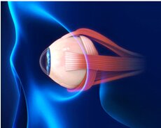
The orbit is a protective bony socket in which the eye rests. The eye is connected to six extraocular muscles in the orbit. These muscles allow the eye to be rotated as well as moved up and down and side to side.
The sclera, the white portion of the eye, is where the extraocular muscles are connected. Nearly the whole surface of the eyeball is covered by this robust layer of tissue.
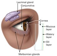
A transparent membrane known as the conjunctiva covers the exterior of the eye and the interior surface of the eyelids.
Tears have three layers and lubricate the eye. The tear film is the collective name for these three layers. The conjunctiva produces the mucous layer. The lacrimal gland produces the watery portion of tears. The lacrimal gland of the eye is located in the orbit, behind the outer brow, away from the nose. The oil that is a component of the tear film is produced by the meibomian gland. The tear duct allows tears to exit the eye.
The cornea, the transparent, dome-shaped front part of the eye, is where light is focused into the eye.
The anterior chamber is the region behind the cornea that contains fluid. The substance is known as aqueous humor. Aqueous humor is continuously produced by the eye. Aqueous humor also drains from the eye in a region known as the drainage angle in order to maintain a consistent eye pressure.
The iris, which is the colored portion of the eye, and the pupil, a dark hole in the middle, are located behind the anterior chamber. The amount of light entering the rear of the eye is controlled by muscles in the iris that either dilate (widen) or constrict (narrow) the pupil.
The lens is located directly behind the pupil. Light is directed toward the retina by the lens. To aid the eye in focusing on close-up objects, the lens adapts its shape. The lens capsule is suspended from the eye wall by a network of tiny fibers known as zonules. The lens capsule, which protects the lens when it is removed during cataract surgery, surrounds the lens. Some types of intraocular lens replacements are placed inside the capsule, where the natural lens once resided.
The cornea and the lens both contribute significantly to our ability to see clearly by aiding in the focus of light as it enters the eye. In actuality, the cornea and lens together account for 70% of the eye’s focusing power.
Between the lens and the retina is the vitreous cavity. The cavity is filled with vitreous humor, a jelly-like material.
The retina, the light-sensitive tissue lining the back of the eye, receives light that is directed into the eye by the cornea and lens after passing through the vitreous.
Our finely tuned, central vision is provided by the macula, a small but extremely specialized region of the retina. Our peripheral (side) vision is provided by the peripheral retina, which is the other portion of the retina.
Photoreceptors are unique cells found in the retina. Light is converted into energy by these cells, which is then sent to the brain. Rods and cones are the two different types of photoreceptors. Rods are responsible for seeing in the dark and for night vision. Cones offer center (detail) vision and color perception.
Through the optic nerve, the retina transmits electrical impulses of light to the brain. The visual cortex, the area of the brain that controls our vision, receives these impulses from the millions of nerve fibers that make up the optic nerve.
At The Eye Center- Dr. Mahnaz Naveed Shah & Associates our team of eight ophthalmology subspecialists/ eye specialists, eye surgeons who are considered amongst the very best eye specialists in Karachi and in Pakistan, have the diagnostic and treatment capabilities to treat from the simplest to the most complex patients. We work hard to provide our patients with the best possible medical and surgical eye care, in a state of the art purpose built eye care facility. We offer the entire array of medical, laser and surgical treatments to help provide patients the best possible care in the most efficient, safe and ethical manner.
If you need an appointment, please contact us at 03041119544 during our working hours or leave us a WhatsApp message at +923028291799 and someone will connect with you. Walk-in appointments are also available for emergencies. We can also be reached through our web portal on www.surgicaleyecenter.org